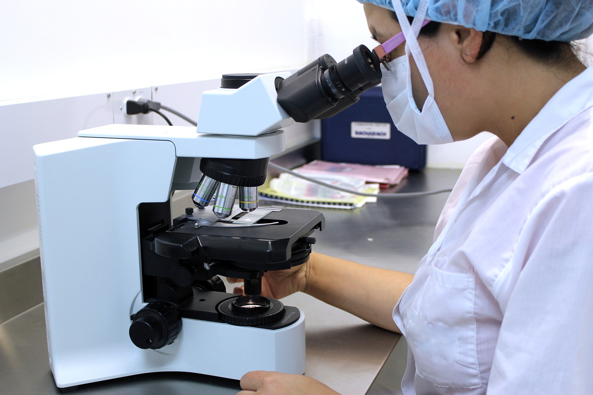
An important scientific lab instrument to observe and analyze the micro-objects even though cells. In simple words, we use a microscope to see objects that cannot be seen through the naked eye.
A Dutch scientist, Zacharias Janssen is the first scientist who laid the foundation of the microscope by inventing the telescope or the true compound microscope.
Robert Hooke is also known as the founder of the simple microscope to examine the cork minutely. That was a simple microscope with one or two lenses, an object holder, and an eyepiece. This simple microscope magnified the object at maximum level but did not use to study the anatomy of the object. Robert hook uses the lamp to illuminate the microscope.
After these discoveries, scientists continued to work on it. They upgrade the microscope at an astonishing level. Now a day, unique microscopes are used to see the external and external structure of the object and also use to see the function of the organelles.
Today, you see different types of microscope used at institutional levels and different labs according to their use

- Simple microscope
- Compound microscope
- Electron microscope
- Phase-contrast microscope
- Interference microscope
Simple Microscope
A simple microscope is just used for the magnification of minute objects.
Convex or biconvex lenses are used in the simple microscope. This lens makes the real and erect image of the object. These lenses are adjusted in such manners that make the magnified image of the object.
Compound Microscope
A compound microscope is also used for magnification but its magnification power is more than the simple microscope.
It works in a way that, two convex lenses are used for clear focal lengths. A compound microscope makes an inverted, erect, and magnified image of the object. It can also use in universities or laboratories to see the micro specimens.
Electron Microscope
This microscope was made by M. Knoll and Ruska in 1932 in Germany. In this microscope, he used a source for an electron beam, a condenser lens, and specimen displacing. The electron beam has uniform velocity and the lens that is used to concentrate the beam of electron on the specimen.
Electron microscope further has two types
- TEM stands for transmission electron microscope
- SEM stands for Scanning electron microscope
Both TEM and SEM have their specific use in different fields of science according to their structure.
Phase-Contrast Microscope
This microscope is used to study the anatomy of the cells. In living cells, it shows the action of living cells and observes the changes that occur during mitosis especially changes in the cytoplasm and nucleus. You can also see the effects of various chemicals inside the living organism through the Phase-contrast microscope.
This microscope uses a phase contrast mechanism for the observation that is mentioned in the first paragraph. It makes the contrast image that can easily understand by scientists and students.
Interference Microscope
The interference Microscope is an astonishing invention of scientists in the field of technology. This microscope has made the life of researchers easy. Now they can easily study macromolecules of the body and their function also. Proteins, lipids and nucleic acids, etc are discovered through the interference microscope.
3D interferogram of the specimen is made that has a dark and bright fringe pattern. This microscope is mainly used in advanced research laboratories for innovation in the field of biological science.
