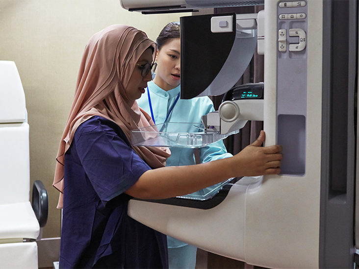
Mammography is a type of breast screening in which the breast must be fixed and squeezed in an X-ray machine designed specifically to study this area to get a clear image.
It’s a simple technique, but it’s also somewhat delicate. Because a safe diagnosis relies on small details, the image quality acquired must be excellent. Fine microcalcifications, for example, that could indicate a severe pathology, could go undetected if technology is misused. Mammography screening clinic Boise takes roughly 10 minutes to complete the exam.
Preparation
- Use no deodorant, powder, perfume, or lotion on the breasts or armpits the day of the exam. These drugs leave behind residues that can taint the outcome.
- Bring all previous exams, including mammograms, breast ultrasounds, and biopsy results, to the mammography screening clinic. The radiologist will be able to generate a more comprehensive comparison report due to this.
- For a few seconds, the gland is compressed, causing a pressure sensation. It is advised that you schedule the checkup after your period to reduce discomfort.
- It’s also a good idea to cut off caffeine (coffee, tea, chocolate, and colas) two weeks or more before the exam. It is recommended that those with a lot of soreness in their breasts take an analgesic 24 hours before and on exam day.
- If there is any suspicion of pregnancy, the mammography, like other radiological procedures, should be postponed unless the treating physician specifies otherwise.
After the exam
Within 24 hours, the outcome will be available. The report was compiled after the examination if the patient requests it, and the radiologist can do so.
Remember to bring any previous tests with you on the day of your visit.
What effect does radiation have on the breasts?
With today’s technology, the amount of radiation received by the breast in an adequately performed examination is small; but, as with any exam that employs ionizing radiation, mammography has biological consequences on the breast; therefore, only what is necessary should be done.
The radiation received by the mammary gland during exposure is reduced by roughly 20% when digital mammography is used.
What’s the difference between a mammogram and an ultrasound?
They’re meant to be used in tandem. Mammography aids in the detection and analysis of microcalcifications. In contrast, ultrasound aids in the search for nodules in dense breasts, the study of internal tumor characteristics, the vascularization of lesions, and the correction of differential diagnoses using color Doppler and other vital data.
Should all women get a breast ultrasound in addition to their mammogram in Boise?
When a mammogram reveals an aberrant picture (most commonly a nodule, distortion, asymmetric densification, or specific microcalcifications), a guided ultrasound is used to diagnose the lesion’s etiology.
Sometimes mammography reveals no abnormalities, only a higher density of the tissue. The identification of cancer is easier in fatty breasts (types ACR 1 and 2) and more complex in dense breasts (types ACR 3 and 4). (type ACR 3 and 4). The latter requires a supplemental study with ultrasonography to increase identification, whereas adipose (ACR 1) and somewhat dense (ACR 2) breasts may not need it.
Indications
- Breast cancer can be detected early.
- Benign breast cancers are studied.
- Examined before surgery (before all types of plastic surgery).