The human shoulder, an intricate symphony of bones, muscles, and ligaments, holds the key to our remarkable range of motion. Whether we’re reaching for the top shelf or throwing a winning pitch, the shoulder joint is at the heart of these movements. But, hidden beneath this seemingly effortless grace lies a world of complexity and vulnerability. Enter the MRI, a groundbreaking technology that unveils the secrets of the shoulder like never before. In this exploration, we embark on a journey through the realm of MRI shoulder imaging, uncovering the insights it offers and its pivotal role in reshaping the landscape of shoulder care.
Understanding the Shoulder Joint
Imagine the shoulder as a harmonious dance between bones, tendons, muscles, and ligaments—a choreography that allows us to embrace, lift, and perform countless feats. This joint’s anatomy reads like poetry: the ball-shaped head of the humerus fitting into the shallow socket of the scapula. The intricate network of tendons and muscles, including the legendary rotator cuff, orchestrates movement, stability, and strength. Yet, this complexity comes at a cost; the shoulder is prone to injuries, and its susceptibility often mirrors its versatility.
MRI Technology: A Glimpse Inside
Picture a world where diagnosis is achieved without incisions or radiation. This is the magic of MRI technology. By harnessing the power of magnetic fields and radio waves, MRI scans create a detailed tapestry of the body’s internal structures. Unlike its radiation-emitting counterparts, MRI is a gentle giant, offering images of unparalleled detail. This imaging revolution allows us to venture into the heart of the shoulder, mapping its intricate anatomy and unraveling the mysteries of its ailments.
Indications for MRI Shoulder Imaging
When the shoulder rebels with pain, stiffness, or diminished function, it’s often a sign of underlying issues that demand attention. Conditions such as rotator cuff tears, labral tears, shoulder impingement, arthritis, and bursitis are just a few of the culprits that can dampen the shoulder’s symphony. This is where MRI shoulder imaging takes center stage. The non-invasive nature of MRI scans shines a spotlight on the inner workings of the shoulder, revealing the hidden intricacies that fuel discomfort and disruption.
Insights from MRI Shoulder Imaging
Step into the realm of MRI shoulder imaging, and you step into a world of revelations. These scans unveil more than just images; they unfold stories of the shoulder’s health. Consider the insights they provide:
- Soft Tissue Evaluation: MRI delves into the fabric of the shoulder, examining muscles, tendons, ligaments, and cartilage for signs of tears, inflammation, and degeneration.
- Rotator Cuff Assessment: The rotator cuff’s integrity is paramount. MRI scans detect partial or full-thickness tears, offering a roadmap for targeted interventions.
- Labral Examination: The labrum, a ring of cartilage around the shoulder socket, is prone to tears. MRI unveils these subtleties, guiding treatment strategies.
- Bursa and Joint Space: The bursa sac’s inflammation and joint space’s condition hold significant diagnostic clues. MRI unravels these narratives, aiding in arthritis detection.
- Blood Flow and Inflammation: Inflammation and compromised circulation don’t escape MRI’s gaze. These scans spotlight areas of concern that might otherwise remain hidden.
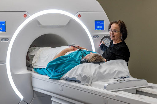
The MRI Shoulder Imaging Process
As patients step into the world of MRI shoulder imaging, the experience is akin to embarking on a journey of discovery. After a few pre-scan instructions—shedding metal objects and donning appropriate attire—the MRI machine becomes a portal into the shoulder’s inner sanctum. Remaining still during the scan might seem challenging, but the payoff is immeasurable—a window into the intricate world of the shoulder’s architecture.
Interpreting MRI Results
While the images might be captivating, their true significance requires the trained eye of medical experts. Radiologists and orthopedic specialists become the narrators of these visual tales, deciphering the nuances that hold the key to diagnosis. A tear, a faint shadow, or an inflammation hotspot—these are the breadcrumbs that lead to tailored treatment paths. It’s through these experts’ interpretation that patients gain insights into their shoulder’s health.
Treatment and Care Pathways
The transformational power of MRI shoulder imaging extends beyond diagnosis; it’s a linchpin for treatment strategies. Armed with the insights gleaned from MRI scans, healthcare providers craft personalized care pathways. From physical therapy to medications, injections to surgical options, MRI data guides the way. It’s a journey toward healing, one illuminated by the precision of modern medical marvels.
Advancements in MRI Shoulder Imaging
As technology marches forward, MRI shoulder imaging evolves as well. Technological strides enhance the accuracy and efficiency of scans, ushering in a new era of diagnostic excellence. Among these innovations stands functional MRI (fMRI), a visionary approach that peers into neural activity related to shoulder movement and pain perception. This pioneering horizon promises to deepen our understanding of the shoulder’s mysteries.
Conclusion
In the enigmatic expanse of the human shoulder lies a symphony of movement, strength, and vulnerability. When pain or discomfort strikes, MRI shoulder imaging emerges as a guiding light, illuminating the path to relief. With each scan, the curtain lifts on the intricate narratives of muscles, tendons, and cartilage. Upright MRI of Deerfield stands at the forefront of this transformative journey, harnessing the power of MRI to unveil the shoulder’s secrets. So, to all those navigating the labyrinth of shoulder discomfort, remember that within the magnetic embrace of MRI lies the key to unraveled mysteries and restored vitality.