Depending on the patient’s condition, vascular access can be performed using several different methods. These include an AV graft, a Fistula, a Catheter, and Ultrasound. However, some patients have poor veins and have difficulty finding a suitable vein. In such cases, an unusual venous entry site may be needed.
AV graft
AV grafts are vascular access solutions made from artificial segments that are inserted between a patient’s artery and vein. They can be placed in the upper arm, thigh, or lower leg. A variety of synthetic materials and configurations are used to create the graft.
When AV grafts are placed, the patient is able to use the graft two to three weeks after surgery. However, they are prone to problems like blood clotting and infection. Nevertheless, well-maintained grafts can last for years.
Fistula
The success of a fistula for vascular access depends on how it is performed and how it is maintained. In general, the fistula can be used for several months after surgery. However, fistulas can fail prematurely or immediately. This is particularly true for elderly or diabetic patients.
Approximately 3% of patients require a second surgery to correct the fistula and restore blood flow to the affected hand. It is usually performed as an outpatient procedure. The procedure takes about one to two hours, and you can usually return home the same day.
Catheter
A catheter for vascular access is a vascular access device. It comprises a cannula 2 with at least a first lumen 21 and at least one first connection tube 3. The catheter also has a single connection means 4, which is overmolded on at least one end of the cannula. The connection allows fluid and mechanical communication between the catheter and the vessel.
Patients with vascular disease are more likely to receive a catheter for vascular access than those without. However, patients with vascular disease are at increased risk for catheter-related mortality. Patients with vascular disease are also more likely to have other comorbid conditions. However, even after adjusting for comorbid conditions, the risk of death associated with catheter use was not reduced.
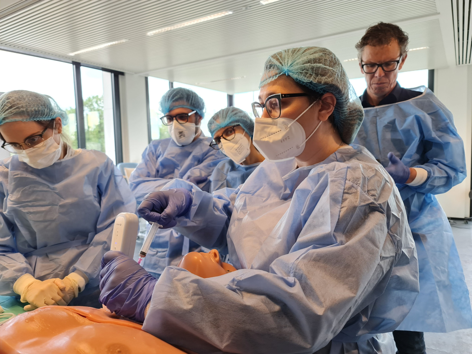
Ultrasound
Ultrasound guidance during vascular access procedures has proven to be a valuable tool for surgeons, enabling them to visualize the underlying structure and enhance the success of the procedure. The technique has also been shown to reduce the need for central lines, resulting in a shorter procedure time and improved patient satisfaction.
Ultrasound is used to guide the insertion of a catheter during central venous access procedures. Traditional sites for central venous access include the internal jugular vein, subclavian vein, and femoral vein. However, peripherally inserted central catheters have gained in popularity over the past decade, particularly in patients requiring prolonged central venous access. Ultrasound guidance is also used to assist in the placement of arterial catheters and peripheral intravenous lines.
Thrombectomy
The procedure is performed through a small incision, and the vascular specialist will insert a catheter into the affected vein. A small balloon attached to the tip of the catheter inflates as the doctor moves it further into the vein, dissolving the remaining clot. The catheter is then withdrawn from the vein, taking the clot with it. The patient will typically be able to go home the same day.
In this systematic review, we examined randomized studies of thrombectomy with vascular access. We searched the Medline database for papers published in English. Our search terms included vascular access, thrombosis, and hemodialysis. We also included large population-based studies, randomized trials, and meta-analyses of the available studies. We excluded studies with small numbers of patients. Additionally, we analyzed studies involving arteriovenous grafts separately from those involving arteriovenous fistulas.