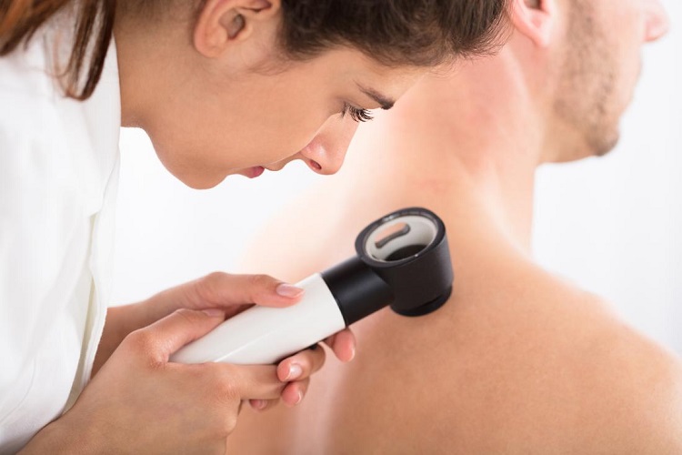
A qualified doctor or nurse should use a standard computer dermoscopic camera and skin mapping technology to perform melanoma examinations through digital photography. High zoom and high light intensity are used in digital dermoscopy to illuminate the sub-surface characteristics of skin lesions that are not visible to the naked eyes It is especially well suited to the detection of skin cancer though it can also be used to diagnose non-melanoma lesions such as basal cell carcinomas. Remote diagnosis, earlier skin cancer identification, skin check, and preventive medicine may all get over here the benefit from digital imaging.
Diagnostic Accuracy
Although modern dermoscopic techniques can provide reasonable diagnostic accuracy digitally ct scan blacktown, dermoscopy can improve skin cancer diagnosis accuracy. During a digital skin check, photographs of all lesions that meet specific conditions that could suggest malignancy are taken. A standard image photograph of a lesion is usually taken first, followed by a high-resolution dermoscopic image. It is also possible to record information about a lesion, like itchiness or perceived development. Digital dermoscopy can assist health professionals to diagnose melanoma and decrease the requirement for needless excision of benign lesions by combining these specifics in an online database. This technique may also decrease the need for biopsies by medical professionals.
Advantages of Digital Record-Keeping
One of the most important advantages of digital dermoscopy is that if a digital document is created, it can be likened to pictures captured throughout subsequent consultations. This allows medical professionals to spot improvements in the skin soon on and diagnose melanomas until they develop an issue. This is particularly beneficial for patients with a huge number of moles (50 or more) or atypical moles (5 or more), as it can be hard for doctors to monitor improvements in such large numbers. It’s also beneficial for patients who are at high risk of developing melanoma skin cancer, as many skin cancers develop from earlier unchecked skin and spread quickly. Patients who have digital imaging performed are routinely reviewed at least once a year.
Applications in Telemedicine
A physician or dermatologist may review digital images on-site, but they’re more commonly utilized in telemedicine, where a doctor or dermatologist reviews the photographs remotely. Patients in isolated places who do not have access to expert doctors may benefit from this. A dermatologist or specialist doctor would send a referral to a local family physician after diagnosing each lesion, recognizing any lesions of importance, and describing any proposed treatment. Digital dermoscopy could also help treatment centers with high patient volumes by allowing a nurse or common practitioner to accomplish the time-consuming task of a photograph capturing while a professional doctor or dermatology St George Utah performs the examination later. The expert will review the images of a large number of patients in a short period of time by concentrating on the diagnosis.
Conclusion:- On a daily basis, a detailed inspection of the person must be performed. This is the most effective way to avoid skin cancer. Moles must be checked for their shape, color, and size if they appear. A doctor’s assistance would be preferable.